In vitro and in vivo imaging of nanoparticle-conjugated antibodies with magnetic particle imaging
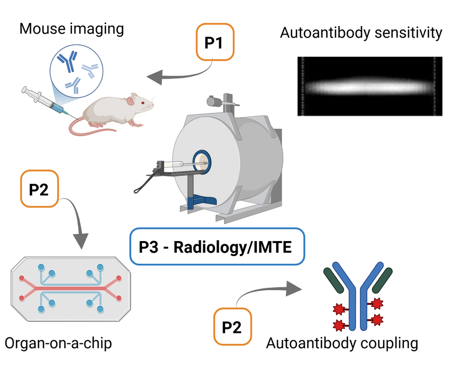
Project 3 aims to establish the new technology Magnetic Particle Imaging (MPI) as a tool to examine autoantibody binding in living organisms. It will be performed by the Department of Radiology together with the Institute of Medical Engineering (IMT)/Fraunhofer Research Institution for Individualized and Cell-based Engineering (IMTE). MPI is a novel imaging technique that utilizes superparamagnetic iron oxide nanoparticles (SPIONs) as tracers. The core principle of MPI involves applying magnetic fields that selectively excite these nanoparticles, allowing for high spatial and temporal resolution imaging without ionizing radiation. MPI is particularly sensitive because it detects only particle signals and not signals from anatomical structures. A unique feature of MPI is its ability to provide particle contrast, known as Multi-Contrast-MPI (MC-MPI). This allows for the simultaneous visualization of different types or characteristics of nanoparticles within the same imaging session. The successful application of MC-MPI could exemplarily be shown for different types of particles, different particle temperatures as well as the binding state in cellular uptake. For the proposed project MC-MPI offers exceptional potential in visualizing antibodies. SPIONs are chemically conjugated with specific antibodies together with P2. After testing various conditions, the antibody-nanoparticle conjugates are injected into the biological system, such as animal models or cell cultures in collaboration with P1 and P2, respectively and interaction between cells and organs will be visualized.

Principle Investigator

PD Dr. Franz Wegner

Thom van Ommeren

Dr. Ing. Mandy Ahlborg