OCT-A-based evaluation of retinal vasculature in mouse and human
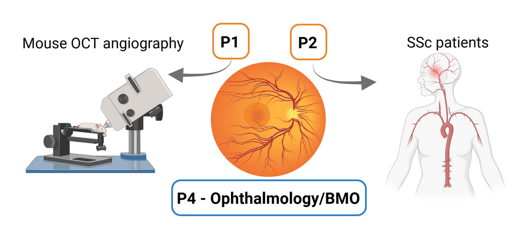
Project 4 aims to examine the eye vasculature in autoantibody-mediated diseases using optical coherence tomography (OCT) in humans and mice. It will be performed by the Department of Ophthalmology and the Institute of Biomedical Optics. OCT angiography is a method that is routinely used in ophthalmology to visualize ocular blood vessels. In addition to structural information, OCT angiography can be used to determine blood flow in the retinal and choroidal vasculature which allows us to determine functional impairments in autoimmune diseases. Together with P2 SSc patients will be examined and correlations to the findings of P2 in neurological assessments and markers of endothelial dysfunction will be drawn to identify targets that can be used to treat neurological symptoms in autoimmune diseases. OCT angiography can visualize vascular effects in neuronal tissue (retina) in humans and could play an important role in diagnosis and treatment control. In addition, OCT angiography will be established in mice to examine the effects of the AT1R-AAB on the ocular blood flow in animals. This will be performed in close collaboration with P1 and allow us to identify similar pathologies between the retinal and the brain vasculature and between mice and humans. In future studies, pharmacological and genetic interventions will be used in mice to resolve the mechanisms by which autoantibodies might affect the neurovasculature.

Principle Investigator

Prof. Dr. Yoko Miura

Prof. Dr. Gereon Hüttmann